A growing number of MRI sequences are validated for neuroscience researchers.
Anatomical acquisitions
These are 3D T1 or T2 weighted or quantitative acquisitions that can be used to conduct morphometry studies or to be used as an intermediate registration target in functional or diffusion MRI, or for crossover with other modalities such as Magnetic Encephalography (MEG) or Transcranial Magnetic Stimulation (TMS)…
Advanced sequences: several state-of-the-art possibilities have recently been added to the MRI Center’s catalog thanks to the collaboration and sharing of different international research centers:
– multi-echo for MPRAGE or MP2RAGE sequences: possibility to combine the echo for better SNR or for additional quantitative measures (T2* maps or to get different contrasts..)
– prospective motion correction: by vNav echo navigator (T1w-MPRAGE and T2w-SPACE images)
– retrospective motion correction: by echo navigator FatNav (MP2RAGE images only)
– ultra-fast sequences using Compressed Sensing: MPRAGE, MP2RAGE, MEMPRAGE, MEMP2RAGE, …
– CAIPIRINHA acceleration for the SPACE sequence (T2 contrast) and integrated iterative denoising
T1 weighting
MPRAGE sequence
Resolution of 1mm isotopic: TR/TI/TE= 2400/900/2.98 ms, Parallel acceleration (GRAPPA=3), flip angle: 8°, bandwidth: 240Hz/pix, Acquisition time: 4 mn 05 s
(Compressed Sensing version (parsimonious sampling): 1 min 51 s )
Resolution of 0.8mm isotropic: TR/TI/TE= 2400/900/2.98 ms, parallel acceleration (GRAPPA=2), flip angle: 9°, bandwidth: 240Hz/pix, Acquisition time: 7 min 02 s
(Compressed Sensing version (parsimonious sampling): 2 min 30 s )
MP2RAGE sequence
Resolution of 1mm isotropic: Acquisition time: 8 min 52 s
(Compressed Sensing version (parsimonious sampling): 4 min 30 s )
T2 weighting
SPACE sequence
Resolution of 1mm isotropic: acquisition time: 2 min 30 s (CAIPIRINHA 4 acceleration)
Resolution of 0.8 mm isotropic: acquisition time: 7 min 58 s (GRAPPA 2 acceleration)
Quantitative T1 imaging
included in the MP2RAGE sequence
Example of anatomical images:
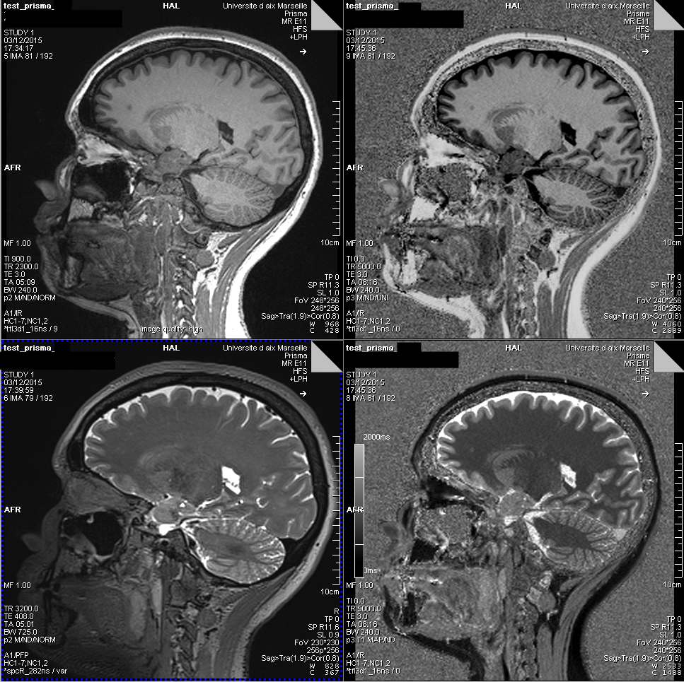
Top Left: MPRAGE T1 weighted image
Top right: MP2RAGE T1-weighted image (free of antenna reception bias)
Bottom left: T2 SPACE weighted image
Bottom right: T1 quantitative image generated by the MP2RAGE sequence.
Functional BOLD images
State of the art functional sequences have been calibrated and validated by the MRI center:
- possibility of multiple accelerations: multiband, parallel acceleration (GRAPPA)
- possibility of multi-echo acquisitions
- SPARSE/Clustered SPARSE acquisitions (Silence/pseudo-silence time for the presentation of auditory stimuli)
- tailored excitation: Zooomit function
- Z-shim: specific shim per slice
- spiral acquisition (to be validated)
- 3D-EPI BOLD acquisitions
Example of protocols:
Time resolution of 1.224 s / spatial resolution of 2.5 mm isotropic: 54 slices, multiband factor = 3, TE=30ms
Time resolution of 0.945 s / spatial resolution of 2.5 mm isotropic: 56 slices, multiband factor = 4, TE=30ms
Time resolution of 0.955 s / spatial resolution of 2 mm isotopic: 60 slices, multiband factor = 5, TE=35ms
Time resolution of 1.386 s / spatial resolution of 2 mm isotopic: 72 slices, multiband factor = 4, TE=33,4ms
Diffusion images
As for the functional MRI sequences, different state-of-the-art sequences are available.
– multiple acceleration possibilities: multiband, parallel acceleration (GRAPPA)
– targeted excitation: Zooomit function
Several protocols are calibrated on the Prisma, with more or less scattering directions, and the choice to repeat all these directions with reverse phase encoding or only some b0 in reverse encoding. All this will have an impact on the acquisition time.
Example of a protocol:
1.8mm iso resolution, 7x b0, 6x b300, 32 b1000 and 64b2000. The 102 directions are unique and optimized inter-shell and intra-shell (Caruyer method). These 102 directions are repeated in AP and PA to use the FSL eddy least-square resampling method.
–> Acquisition of 11 minutes in total.