The stimulation bench of the MRI Center is a hardware and software environment allowing researchers in neuro-imaging, integrative neurosciences, cognitive or clinical psychology, to interact in the most complete and precise way possible with subjects (human or PNH) installed in the MRI imager, in order to make possible the smooth running of experiments in cerebral functional MRI.
This development of instrumental integration can be represented formally and functionally as a three-layer architecture, essentially involving the three fields of computer science, electronics and mechanics
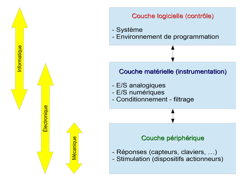
- The peripheral layer is the most visible to users. It concerns all the devices for collecting behavioural data and stimulating the subject. In a word, it concerns the devices for interaction and interfacing with the subject.
- The hardware layer is clearly the least visible: it concerns all the analog, digital and video input/output cards, as well as the conditioning devices (filtering, amplification, isolation, etc.).
- The software layer allows to orchestrate the bench and to control in real time the whole chain synchronized with the MRI system. It obviously includes the programming environment (LabVIEW in this case) but also the operating system. This layer also provides the interface user-friendly and ergonomic with the experimenter (Man-Machine Interface, MMI).
The balance, robustness and suitability of these three layers is crucial for the proper functioning of the entire environment, according to the different levels of functionality and requirements.
The entire modular bench is shown in the following figure. The three layers are shown in light yellow, orange and blue. The green modules are the visible and usable part for the users (researchers, engineers, students).
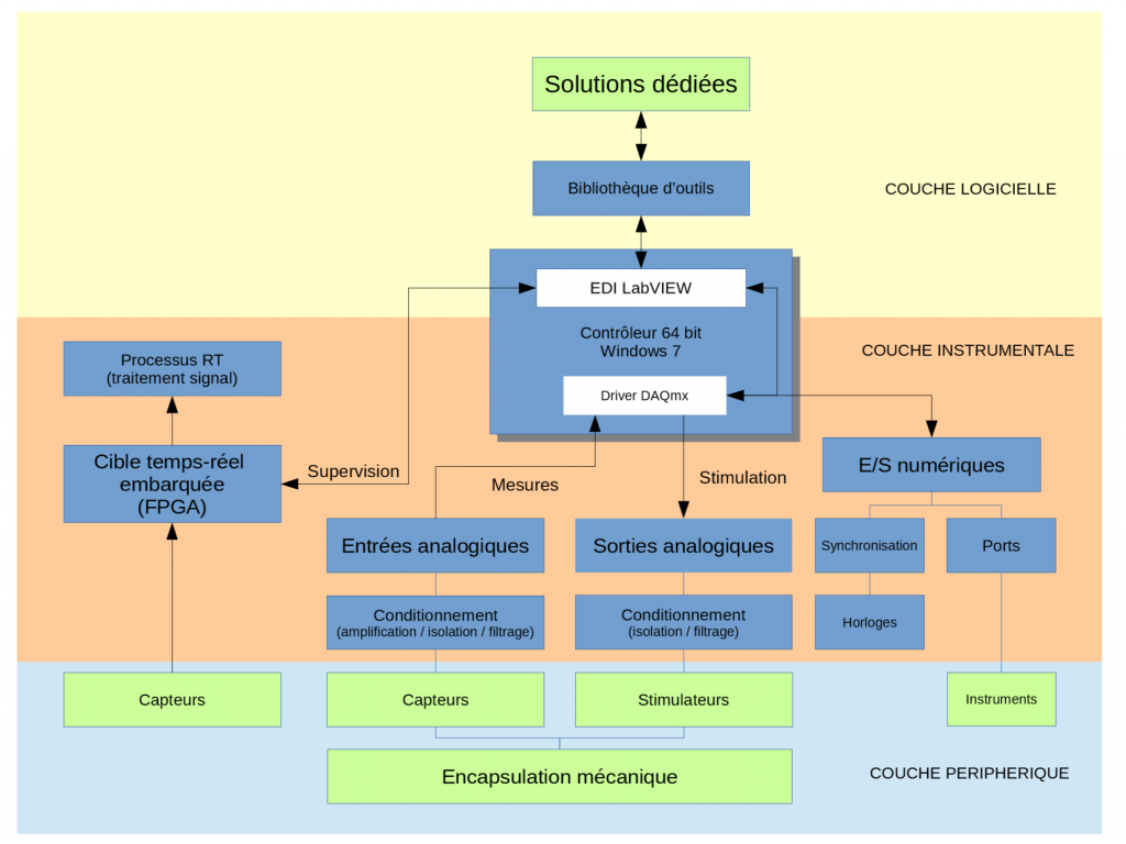
The three layers are detailed below.
– Stimulators
From a material point of view, the main stimulation peripherals developed cover a wide range of perceptive inputs:
- Visual stimulation: LCD video-projector, diode arrays, programmable occlusion glasses
- Auditory stimulation: pneumatic, piezoelectric, electrodynamic systems
- Skin stimulation: piezoelectric vibrators, electrical stimulation
- Olfactory stimulation: pneumatic bench for odour diffusion, synchronised with respiration
- Proprioceptive stimulation: pressure-controlled pneumatic vibrators
– Collection of behavioral responses
Similarly, the various response collection devices also tend to cover the widest possible panel, depending on the needs of the teams:
- Multiple-choice behavioural responses: ergonomic 5-button keypads (right hand, left hand), answer buttons. Precise measurement of reaction times using electronic counters.
- Visual Behavior: EyeLink 1000 Eye-Tracking System
- Motor responses: non-magnetic joystick, optical trackball, joint movements, optical movement sensor, analogue force sensor
- Voice response: MRI-compatible optical audio recording system with real-time amplification and noise cancellation
- Physiological responses: heart rate (plethysmography), respiration, electro-dermal response, swallowing, ElectroMyograGraphy (EMG)
- Graphic tablet dedicated to writing or pointing tasks.
All of these peripherals (stimulators or response sensors) are either developed within our platform or in partnership with local players in our network of expertise (engineering schools in the Aix-Marseille area), or purchased from manufacturers, provided that they are perfectly mastered and can be upgraded within our platform.
The instrumental layer is the heart of the stimulation bench. It is articulated around National Instruments materials. This choice is the fruit of a partnership initiated more than 10 years ago and which is anchored in a local, institutional and private network of skills (notably the Provence School of Microelectronics) which enables reciprocal expertise to be shared and numerous materials to be tested in order to validate the prototypes before final purchases.
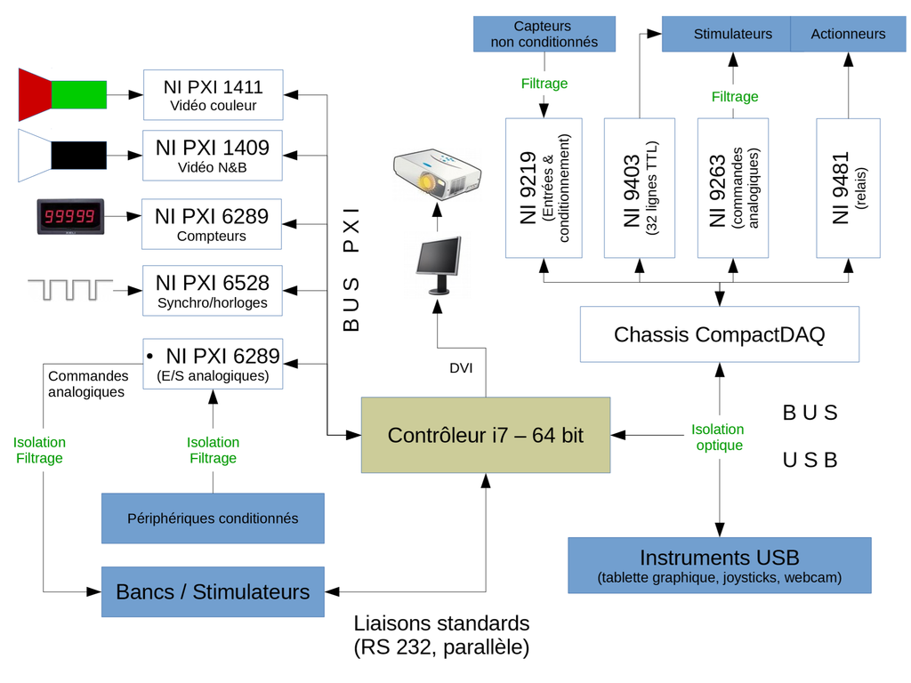
The “Instrumentation” layer and the references of the main components are illustrated above.
The selected architecture is based on the complementarity of the PXI bus (for analog and digital controls, video acquisition and synchronization modules) and CompactDAQ chassis for conditioned I/O (force sensors, resistive sensors, …). All of these devices are addressed by a common driver (DAQmx) which guarantees the portability and scalability of all the modules and is programmed under LabVIEW.
The RS-232 and USB ports are also used in order to allow the integration of more standard instruments (serial actuators, graphic tablet, …).
Finally, a very particular care is given to the Electromagnetic Compatibility of Systems (EMCS): indeed it is necessary to limit all sources of HF or LF electronic noise, especially in an environment that multiplies references (Faraday cage, ground, floating references, power supplies …). We have therefore set up all the tools to limit ground loops and common mode effects using tools adapted to each component: optical insulation, decouplers, converters, power supplies with reinforced insulation, filtering.
All these precautions ensure that all stimulators and sensors can be used in complete safety and with the guarantee of generating and recording the cleanest possible signals.
The entire bench is fully programmed using National Instruments’ Integrated Development Environment (IDE): LabVIEW.
The modularity of LabVIEW, associated with the formalism of data flow programming specific to the G language is perfectly adapted to the architecture of the bench and to our functional constraints.
We have developed a complete library gathering all the functions related to our environment… More than 130 VIs (Virtual Instruments: software modules) have been programmed and grouped by functionalities (management, stimulation, response, backup, synchronization, simulation, I/O, regulation, etc.).
The applications dedicated to each study are developed from a common framework, but each study is the subject of a program with its own GUI, options and input/output data. This program is fully functional in our platform, coupled with the MRI system, but also in “test” mode: in the latter case, the MRI synchronisation is emulated and the researchers can test the protocols (stimuli, time course, behavioural performance) on a simple PC and, if necessary, train the subjects for the protocol in question, with an environment similar to that of the platform.
